Home

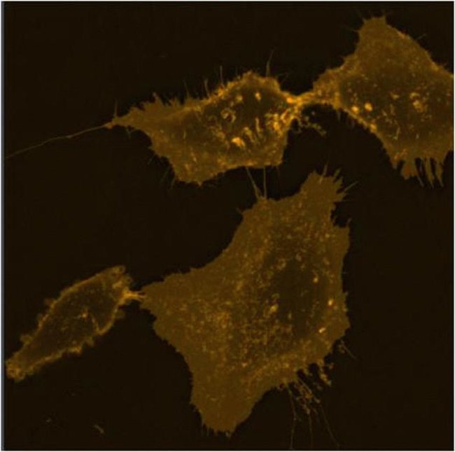
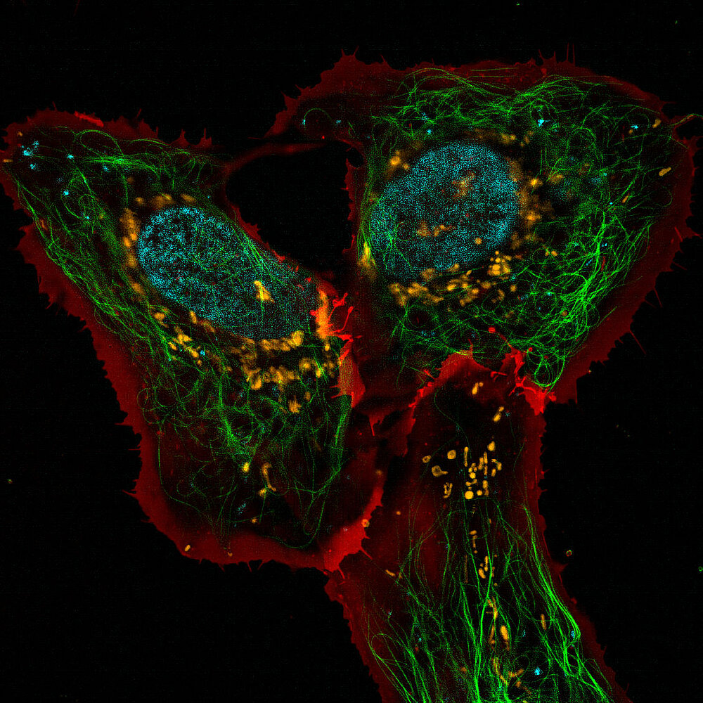
cell mask orange

Cell Mask Orange Plasma Membrane Stain fluorescent microscope |  Biocompare.com Kit/Reagent Review
Cell Mask Orange Plasma Membrane Stain fluorescent microscope | Biocompare.com Kit/Reagent Review

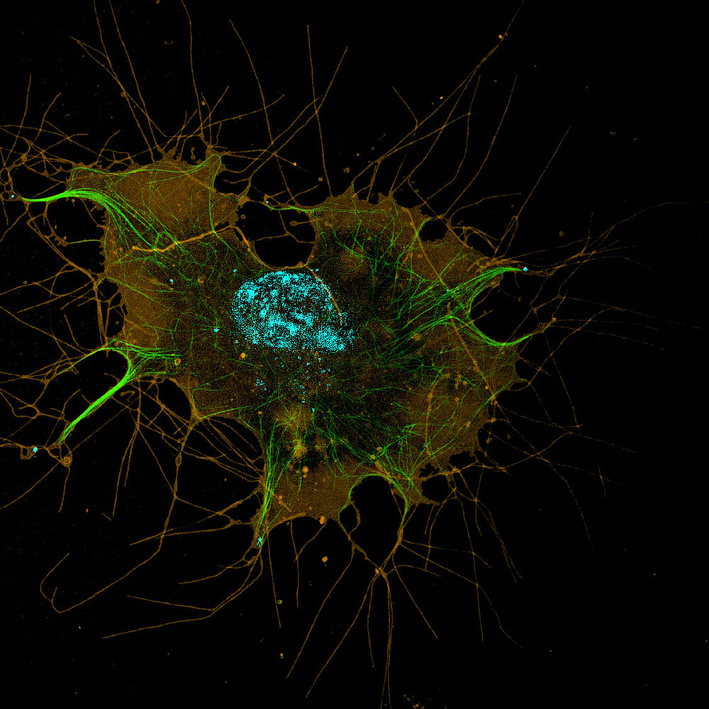
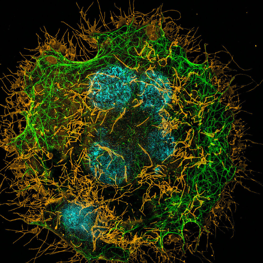
SIM - Chair of Biotechnology

a, d) Structure of (a) NVP 1 and (d) NVP 2. (b, e) Fluorescent images... |  Download Scientific Diagram
a, d) Structure of (a) NVP 1 and (d) NVP 2. (b, e) Fluorescent images... | Download Scientific Diagram

A–C) 3D-SIM of LSECs stained with Bodipy FL C5 ganglioside GM1, a... |  Download Scientific Diagram
A–C) 3D-SIM of LSECs stained with Bodipy FL C5 ganglioside GM1, a... | Download Scientific Diagram

Confocal microscopy image of membrane labelled (CellMask© Orange) U87... |  Download Scientific Diagram
Confocal microscopy image of membrane labelled (CellMask© Orange) U87... | Download Scientific Diagram

SIM - Chair of Biotechnology

An aryleneethynylene fluorophore for cell membrane staining - ScienceDirect

Ventral stress fibers induce plasma membrane deformation in human  fibroblasts
Ventral stress fibers induce plasma membrane deformation in human fibroblasts

FLIM of CellMask orange labeled GPMVs exposed to SWCNTs. (A) Addition... |  Download Scientific Diagram
FLIM of CellMask orange labeled GPMVs exposed to SWCNTs. (A) Addition... | Download Scientific Diagram

CellMask™ Plasma Membrane Stains

Cell membranes were stained with Cell Mask™ orange plasma membrane, EP... |  Download Scientific Diagram
Cell membranes were stained with Cell Mask™ orange plasma membrane, EP... | Download Scientific Diagram
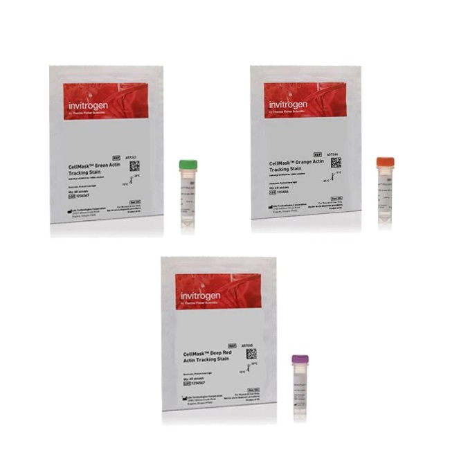
CellMask™ Orange Actin Tracking Stain

LaboShop | Products | Invitrogen™ CellMask™ Actin Tracking Stain Variety  Pack
LaboShop | Products | Invitrogen™ CellMask™ Actin Tracking Stain Variety Pack

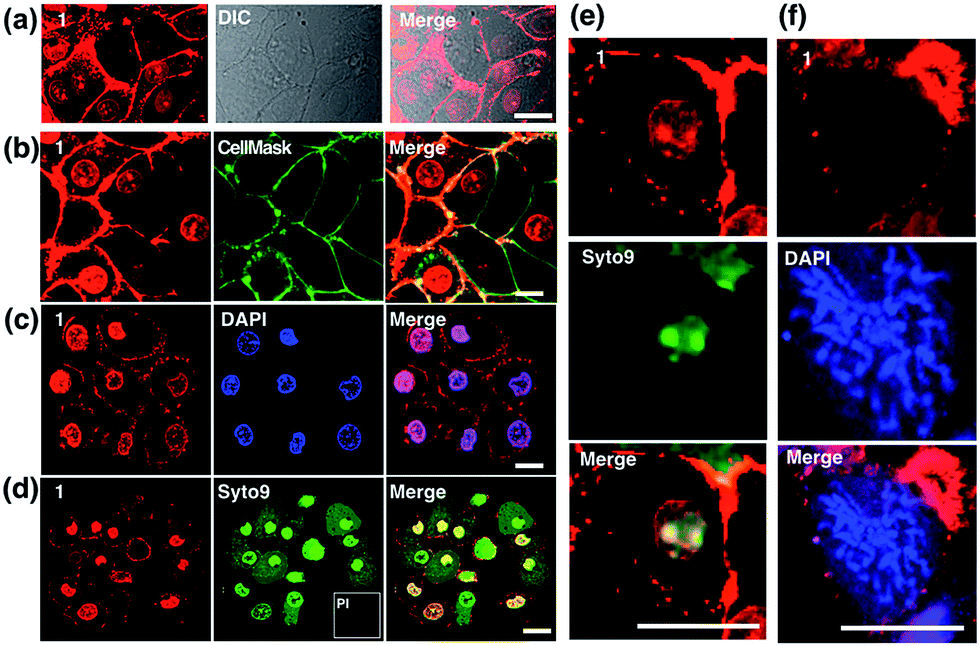
Probe for simultaneous membrane and nucleus labeling in living cells and in  vivo bioimaging using a two-photon absorption water-soluble Zn( ii ) terpy  ... - Chemical Science (RSC Publishing) DOI:10.1039/C6SC02342H
Probe for simultaneous membrane and nucleus labeling in living cells and in vivo bioimaging using a two-photon absorption water-soluble Zn( ii ) terpy ... - Chemical Science (RSC Publishing) DOI:10.1039/C6SC02342H

Hepatocytes Delete Regulatory T Cells by Enclysis, a CD4+ T Cell Engulfment  Process
Hepatocytes Delete Regulatory T Cells by Enclysis, a CD4+ T Cell Engulfment Process

MG-63 cells stained with calcein AM for viable cells, CellMask Orange... |  Download Scientific Diagram
MG-63 cells stained with calcein AM for viable cells, CellMask Orange... | Download Scientific Diagram

CellMask™ Plasma Membrane Stains

SIM - Chair of Biotechnology

Changes in the asymmetric distribution of cholesterol in the plasma  membrane influence streptolysin O pore formation | Scientific Reports
Changes in the asymmetric distribution of cholesterol in the plasma membrane influence streptolysin O pore formation | Scientific Reports

Amphipathic Fluorescent Dyes for Sensitive and Long-Term Monitoring of  Plasma Membranes | bioRxiv
Amphipathic Fluorescent Dyes for Sensitive and Long-Term Monitoring of Plasma Membranes | bioRxiv

Large extracellular vesicles can be characterised by multiplex labelling  using imaging flow cytometry | bioRxiv
Large extracellular vesicles can be characterised by multiplex labelling using imaging flow cytometry | bioRxiv

CellMask™ Plasma Membrane Stains

Muldream Vegan Green Mild Flower Cell Mask – SkinCureAsia

The Relationship between Fenestrations, Sieve Plates and Rafts in Liver  Sinusoidal Endothelial Cells | PLOS ONE
The Relationship between Fenestrations, Sieve Plates and Rafts in Liver Sinusoidal Endothelial Cells | PLOS ONE